
(Header photo, left to right: Emma, George, Isabella, Natasha, and Ellen)
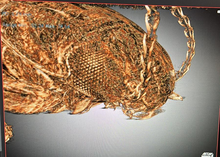
In late June, members of our team and the Quantitative Morphology Group travelled to Melbourne to visit the Australian Synchrotron. There, we used their high-power micro-Computed Tomography (CT) beamline for a continuous 50-hour period to capture the internal and external morphology of microscopic beetles.
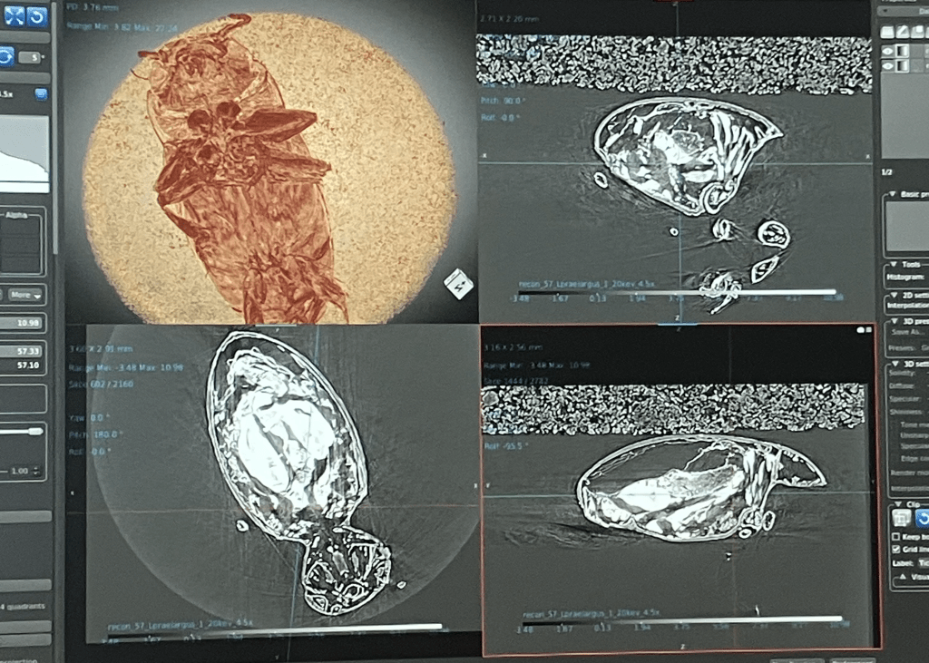
This project aims to investigate trade-offs between visual and chemo-sensory systems in Australian underground water beetles. We conducted high-resolution imaging (min. pixel size = 0.361 µm) of both surface-dwelling and subterranean beetle species, focusing on their brain morphology. We captured detailed 3D models of specimens measuring just 1.5 to 2.5 mm in length, with brain regions as small as 0.2 mm.
Micro-CT is a powerful tool for capturing morphological information in invertebrates. Check out the variety of ways we study invertebrates here!

Leave a comment